By Alice Chou
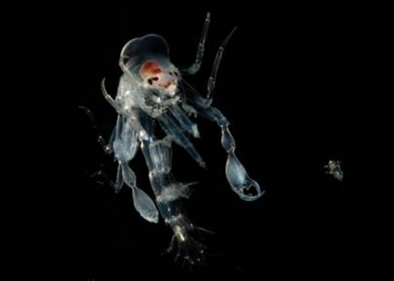
The first time Laura Bagge encountered hyperiid amphipods she looked right through them. While on a research cruise, Bagge, a graduate student researcher at Duke University, was examining deep-sea fish in a bucket. When she reached in to touch the fish, something was in the way. “I ended up hitting this big glass roach-looking thing!” Famed for their unusual and diverse visual systems, hyperiid amphipods are transparent crustaceans that can grow as large as the length of your hand. As part of her research on these glass roaches, she discovered that they possess an unusual anti-reflective coating on their bodies, presumably to evade detection by predators.
Transparency is not entirely unusual in marine environments. Many of the most charismatic ocean dwellers, such as jellyfish and some squid, are also transparent. Because light passes directly through these bodies, no shadows or silhouettes are created to give them away to potential predators.
Unfortunately the strategy is not foolproof, especially for animals with hard shells like hyperiid amphipods. “There are a lot of bioluminescent predators where these animals live,” Bagge explains. “Some fish, like dragonfish, have different photophores that are underneath their eyes that can emit different wavelengths of light.” She likened the function of these bioluminescent organs to shining a flashlight on a window at night. “Even though it’s transparent, that window will be very visible. Any reflections can make [prey] really stand out against the background.”
The presence of anti-reflective surfaces on animals is not a new discovery. In the 1960s, regularly arranged bumps dubbed “nipple arrays” were found on the eye surfaces of some moths and butterflies. These nipple arrays, which have a height of about 200 nanometers, function in much the same way shag carpets in recording studios do. The shag carpet serves as a gradient between the air and the wall, thus dampening incoming waves of sound. Instead of sound, nipple arrays dampen light and reduce the amount that is reflected off an insect. This unique optical adaptation inspired Bagge to look for similar anti-reflective structures on hyperiid amphipods under scanning electron microscopy (SEM).
Conceptually, SEM functions much like light microscopy. However, instead of bouncing light off an object, SEM achieves its image by bouncing electrons off an object’s surface. Its much higher resolution allows researchers to examine minute features of objects, down to measurements on the scale of nanometers. An object is first coated in a very thin layer of gold, which allows reflection of electrons to a sensor, ultimately resulting in a precise and detailed image of surface structures.
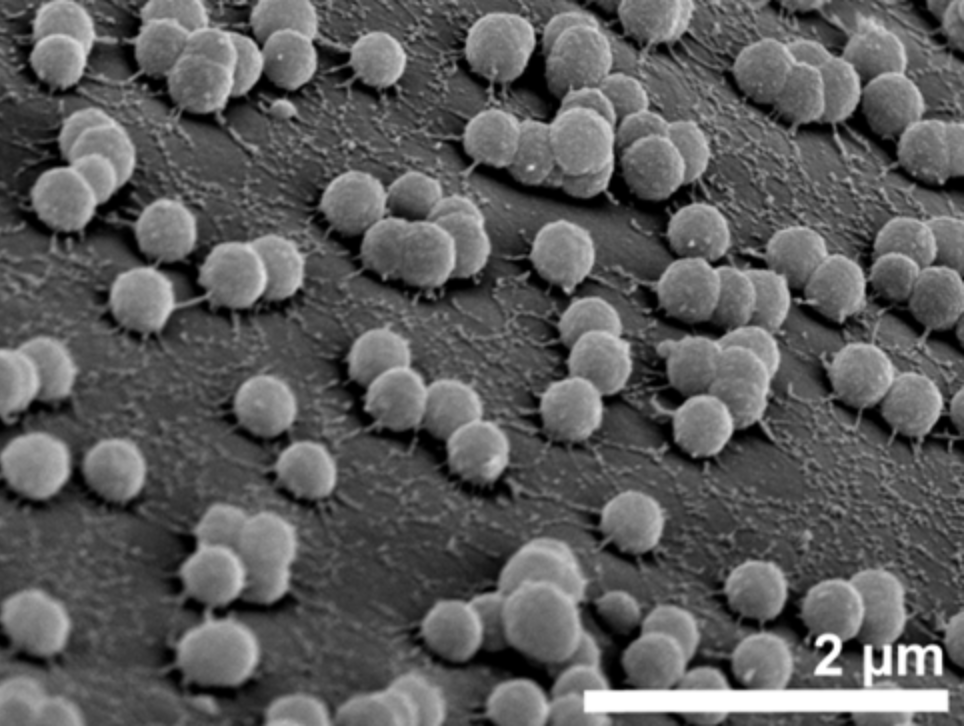
Using this technique, Bagge found tiny spheres on the surface of the amphipods. Unlike nipple arrays, which are physically part of the animal’s exoskeleton, these spheres seem to be distinct from the cuticle—a monolayer. Spheres from different species measured anywhere from 50 to 320 nanometers—which makes them about one thousandth of the average width of human hair. Initially, she thought her results were simply a mistake in sample preparation. “The spheres were so uniform that I thought I had done something wrong with the gold coating!” she laughed, “My day was like, ‘Well. That didn’t work.’” However, additional experiments proved that these unusual results were not the consequence of a procedural error, but may in fact serve an important ecological purpose.
Because the size of the spheres is smaller than the wavelengths of light, they create a form of destructive interference. “[The spheres] act as a gradient in the refractive index. The cuticle surface and the water have different refractive indices. When you move from one medium to another and it’s a flat surface, light is going to bounce right off.” With the monolayer of spheres on hyperiid amphipod cuticles, any light directed at the animals would encounter a gradual change in refractive indices and therefore not be reflected.
Part of what makes Bagge’s discovery so exciting is the possibility that these spheres are living organisms. Upon closer examination of her SEM images, Bagge found that the spheres from specimens of the genus Phronima had small filaments attaching them to the surface of the animal, and that some spheres seemed to be in the process of division. Because the spheres are an order of magnitude smaller than most bacteria, if Bagge’s hunch is correct, these would be the smallest bacteria known to researchers.
To further test her hypothesis that the monolayer is an anti-reflection device, Baggeused mathematical models to determine the amount of reflection off of a cuticle surface given different sizes and distribution densities of nano-spheres. She used the known refractive index of bacteria and found that in all the cases studied, the nano-spheres reduced reflection of the animal’s surface by at least four-fold, and up to two hundred and fifty-fold. Hypothetically, this reduction would be more than enough to evade detection by predators. Her discovery is the first instance of anti-reflection coating on any ocean animal, and the first known use of a monolayer by any animal.
The monolayer of nano-spheres is remarkably similar to thin film anti-reflection technology developed by engineers for use on glasses, windows, and solar panels, suggesting an interesting convergence of animal evolution with human technology.
Her next step is to confirm that the spheres are bacteria by DNA sequencing. However, that hasn’t been without its issues. Immediately after collection, the animals are flash frozen so the monolayer can be painstakingly removed at a later time to be analyzed. “These are watery little bugs. When you’re trying to scrape off the cuticle surface you have this little transparent piece of ice and need to figure out what is what. It’s the worst.”
We should all just be thankful that our terrestrial roaches are visible.